File:Diatoms through the microscope.jpg: Difference between revisions
Jump to navigation
Jump to search
KuroKitten (talk | contribs) (Assorted diatoms as seen through a microscope. These specimens were living between crystals of annual sea ice in McMurdo Sound, Antarctica. Image digitized from original 35mm Ektachrome slide. These tiny phytoplankton are encased within a silicate cell wall. Author: Prof. Gordon T. Taylor, Stony Brook University Permission: Public Domain) |
(No difference)
|
Latest revision as of 09:19, 23 July 2022
Summary
Assorted diatoms as seen through a microscope. These specimens were living between crystals of annual sea ice in McMurdo Sound, Antarctica. Image digitized from original 35mm Ektachrome slide. These tiny phytoplankton are encased within a silicate cell wall.
Author: Prof. Gordon T. Taylor, Stony Brook University
Permission: Public Domain
File history
Click on a date/time to view the file as it appeared at that time.
| Date/Time | Thumbnail | Dimensions | User | Comment | |
|---|---|---|---|---|---|
| current | 09:19, 23 July 2022 | 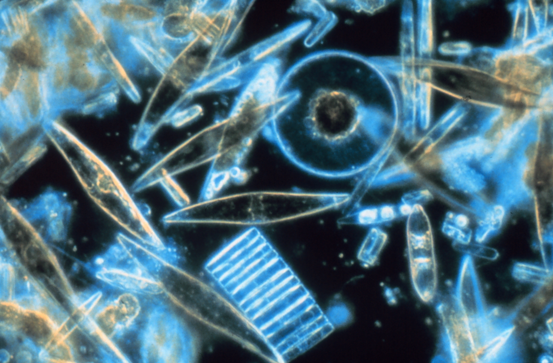 | 1,796 × 1,180 (1.25 MB) | KuroKitten (talk | contribs) | Assorted diatoms as seen through a microscope. These specimens were living between crystals of annual sea ice in McMurdo Sound, Antarctica. Image digitized from original 35mm Ektachrome slide. These tiny phytoplankton are encased within a silicate cell wall. Author: Prof. Gordon T. Taylor, Stony Brook University Permission: Public Domain |
You cannot overwrite this file.
File usage
The following page uses this file: